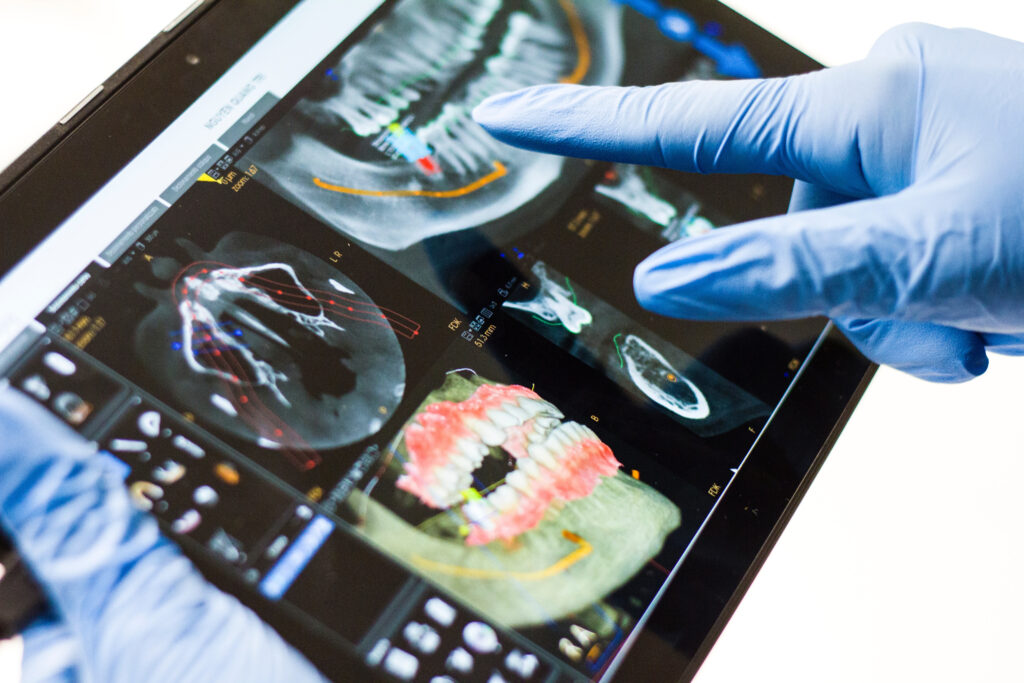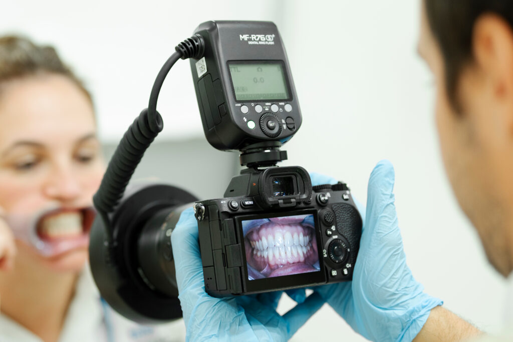
Dental photography or clinical dental photography is an essential part of modern dentistry techniques, used for diagnosis and treatment. This important diagnostic tool helps with many aspects of dentistry, including patient communication, where photos taken of the teeth can help the patient understand their dental situation. It is also crucial for creating treatment plans, keeping a record of a patient’s teeth, and for referrals to specialist clinicians.
Dentists all over the globe are using dental photography more than ever to increase positive patient outcomes and enhance care, so education on this crucial tool is key to many dentists’ career progression.
Read on to learn more about what dental photography is, how it’s used, and how you can enhance your clinical photography skills with the London Dental Institute.
The History of Dental Photography
Dental photography began in the 1950s, as a way for dentists to show their patients what was going on inside their mouths, and help the dentist themselves to diagnose and treat problems. As technology at this time was limited, the best way to take photos of teeth was to use a circular flash attached to the end of a camera lens to provide full illumination inside the mouth. This invention revolutionised the way dentists could communicate with their patients.
In the 1960s and 70s, the popularity of dental photography grew, and the limitations of using 35mm film to take these photos became clear. The need to develop the photos before they could be reviewed would slow down the process of diagnosis, therefore the need for immediate photos became important. The first instant cameras helped this issue immensely, as the camera could take a picture and process it to film inside the camera itself.
In the 1980s, computers took their place in dentistry, and the first foray into computerised photography was undertaken. As computers became more commonplace in medicine, so too did small video cameras with built-in wands that were small enough to fit into a patient’s mouth and display an image on the computer screen. This move towards video rather than pictures was revolutionary, and helped dentists explain diagnoses and treatments to their patients in real time.
These days, almost everyone uses clinical photography video or camera stills to diagnose and treat patients, and the need for good dental photography skills is higher than ever.

What Dental Photography Is Used For
Dental photography is used for many different reasons in everyday practice and specialist areas of dentistry. Taking pictures and videos of the teeth and inside of the mouth benefit both patient and dentist in the following ways:
Diagnosis
Dental photographs provide a baseline reference for a patient’s mouth and teeth at the beginning of treatment. These initial photos can then be referred to when monitoring progress during treatments such as orthodontics, aesthetic procedures, and restorative work.
Treatment
Dentists can utilise clinical photographs to help plan patient treatments following diagnosis, leading to more positive and long-lasting outcomes.
Advance your career by studying for a world-leading postgraduate diploma
Study at the cutting-edge of dental research with curricula taught by global experts and an online schedule that suits you.
Communication
Dental photography helps bridge the communication gap between patients and colleagues when it comes to diagnosis and treatment. For patients and dentists, when a clinician can use a video scope to show the patient their images immediately, they become more aware of the issues in their mouth and the path to treatment. Dental photography is also great for showing progress to patients throughout the treatment process.
Between peers, dental photography helps the communication between clinicians and referral health care providers, for example, if a patient needs to be transferred to a specialist.
Consent & Legality
It is far easier to obtain a higher level of consent from patients to start or continue treatment when dental photography is used, as patients can see the extent of damage or progress for themselves in real-time. These photographic records can also be used and stored to reduce the risk of litigation based on treatment outcomes.
Clinical Skills
Reflecting upon and studying dental photography images enhances the level of patient care one can hope to receive, as well as help the clinician themselves to boost their skills.
Education
Garnering high-quality images as a result of dental photography is important to learn as you work with patients, increasing your skill level every day. Dental photography is also an important component of many postgraduate training programmes, highlighting their importance in everyday practice.
Marketing
Dental photography images can be used in dental portfolios, practice marketing, and research.

Learn More at the LDi
If you’re interested in learning more about clinical photography and how best to utilise this tool to develop your diagnostic and treatment skills, consider enrolling on a postgraduate course with the London Dental Institute.
Diploma In Aesthetic & Restorative Dentistry
Studying for a diploma in Aesthetic & Restorative Dentistry with the LDi is a fantastic way to upgrade your dental photography skills with a faculty of expert tutors and world-class resources. This PG Dip. course is 12 months of remote, flexible learning, so you can boost your skills from anywhere in the world while continuing to work in your dental practice.
This particular course explores dental photography in Unit 2 – Principles of Assessment and Diagnosis. The modules within this unit focus on clinical photography, and students will learn the importance of dental photography for documentation, communication, smile design for aesthetic treatment, and treatment planning for complex cases.
Diploma in Orthodontics & Dentofacial Orthopaedics
Studying for a diploma in Orthodontics & Dentofacial Orthopaedics with the London Dental Institute takes only 12 months of flexible, online study, and will provide students with the skills necessary to diagnose and treat orthodontic issues in patients by utilising dental photography.
The course explores clinical photography in Unit 1 – Principles of Orthodontic Assessment and Diagnosis. The modules within this unit focus on using clinical photography to gain a better understanding of the biology of tooth movements and how to use photos and videos to better understand the needs of patients.
Discover flexible and affordable course options with the London Dental Institute. Speak to an enrolment advisor, or click here to find out more.


